Medical Images in Digital Records: What You Need to Know
Discover the vital role of medical images in digital records. Learn how they enhance clinical decisions and improve patient care.

Medical Images in Digital Records: What You Need to Know
Medical images are defined as diagnostic visual data, including X-rays, CT scans, MRIs, and ultrasounds, that form a core component of electronic health records (EHRs) and directly determine the accuracy of clinical decisions. The role of medical images in digital records extends far beyond simple file storage. These images give clinicians a real-time visual history of a patient’s condition, reduce duplicate testing, and support coordinated care across hospitals, specialists, and primary care providers. The Office of the National Coordinator for Health Information Technology (ONC) has made diagnostic imaging interoperability a top priority in 2026, recognizing that access and exchange across care settings remain a persistent challenge. For patients and providers alike, understanding how these images function within digital health records is no longer optional. It is the foundation of safe, informed care.
How do medical images improve patient safety and diagnostic accuracy?
Medical images embedded in digital records directly reduce clinical errors and improve the speed of diagnosis. When a radiologist or specialist can pull up a prior chest CT alongside a new scan, they can track changes in a lung nodule over time rather than treating each scan in isolation. That longitudinal view is what separates reactive care from genuinely informed clinical decision-making.

The USCDI Diagnostic Imaging Reference standard ties imaging studies to patient records at the point of care, reducing duplicative imaging and improving diagnostic decisions. Fewer repeat scans mean less radiation exposure for patients and lower costs for the healthcare system overall.
Three specific safety benefits stand out:
- Wrong-patient error reduction. Displaying patient photos alongside imaging records in EHRs creates a visual cognitive check that reduces the risk of acting on the wrong patient’s scan.
- Full-fidelity image access. Diagnostic-quality images with complete metadata are required for clinical decisions. Low-resolution previews embedded in some EHR portals are not sufficient for accurate diagnosis.
- Faster specialist consultation. When a primary care physician can share a high-resolution MRI directly with a neurologist through a shared digital record, the referral process shrinks from days to hours.
“Seamless movement of diagnostic images between care teams enables faster, more accurate decisions at lower cost.” — ONC Blog, 2026
Pro Tip: If you are a patient, always request a copy of your imaging study files, not just the written radiology report. The actual image files give any future provider the full picture.
What challenges affect interoperability and image exchange across health systems?
Interoperability is the ability of different health systems to exchange and use medical data without friction. For imaging specifically, this remains one of the hardest problems in digital health. The core issue is that imaging data has historically lived in separate silos from the rest of a patient’s record.
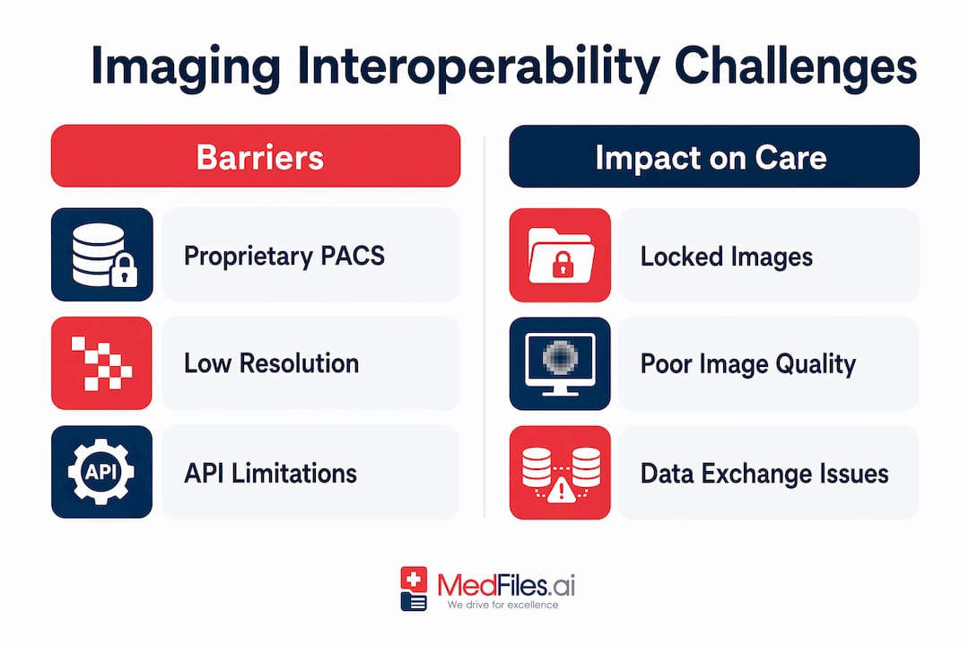
Most hospitals store images in a Picture Archiving and Communication System (PACS) or a Vendor Neutral Archive (VNA). These systems are often disconnected from the EHR, meaning a clinician must log into a separate platform to view images. That extra step creates workflow friction and, in urgent situations, can delay critical decisions.
The four most common interoperability barriers are:
- Proprietary formats and systems. Many imaging vendors use formats that do not communicate with competing platforms, locking images inside a single institution’s infrastructure.
- Inconsistent adoption of standards. DICOM is the global standard for medical image formatting, and HL7 FHIR is the emerging standard for health data exchange. However, fragmented imaging storage and inconsistent format adoption create patient safety risks across the industry.
- Governance gaps. Realizing interoperability benefits depends not only on technical standards but also on governance agreements between organizations hosting imaging systems.
- Resource disparities. Rural and smaller hospitals often lack the IT infrastructure and staff to implement advanced imaging exchange frameworks, widening the gap in care quality.
| Barrier | Impact on care |
|---|---|
| Proprietary PACS systems | Images locked within one institution |
| Low-resolution EHR previews | Clinicians cannot make accurate diagnoses from previews |
| Missing governance agreements | Cross-system image sharing blocked even when technically possible |
| Rural resource gaps | Smaller providers cannot access or send high-quality imaging data |
Imaging interoperability also requires specialized API implementation beyond standard EHR certification, using standards like DICOMweb with delegated authorization to make workflows practical for clinicians. Understanding how patient data affects outcomes helps clarify why solving these barriers matters at every level of care.
What legal rights do patients have to access their medical images?
Patients have a federally protected right to access their medical images. Under HIPAA 45 CFR §164.524, healthcare providers must give patients access to their protected health information, which explicitly includes diagnostic imaging files and radiology reports. Providers must supply electronic copies when the records exist in electronic form, and they have limited authority to charge fees for that access.
What you can request under this right:
- Full imaging study files (DICOM format) from X-rays, CT scans, MRIs, and ultrasounds
- Radiology and pathology reports linked to those images
- Any imaging data used to inform clinical decisions about your care
- Electronic copies sent directly to you or to another provider of your choice
Platforms like those used by Memorial Medical Center now allow patients to access and share imaging records online through secure third-party tools. These systems use Medical Record Numbers and encrypted links, requiring no additional software installation on the receiving end. That model is becoming the standard for patient-centered image access.
Pro Tip: When requesting your imaging files, ask specifically for the DICOM files, not just the printed report. DICOM files are the actual images your doctors use, and any future provider can open them with standard viewing software.
Understanding your health data privacy rights is the first step toward taking control of your imaging records and making sure they follow you across every care setting.
How are new technologies improving medical image retrieval in digital records?
The next frontier in medical data visualization is content-based image retrieval (CBIR). Traditional image search in a PACS or EHR relies on keywords and metadata, meaning a clinician must know exactly what to search for. CBIR flips that model. It uses AI to search imaging archives by visual similarity, returning cases that look like the current patient’s scan rather than cases that share the same text tags.
CBIR systems combined with multimodal AI combine image data with linked radiology reports to return clinically meaningful results. A radiologist reviewing an unusual liver lesion can query the archive for visually similar cases and see how those cases were diagnosed and managed. That is institutional memory made searchable.
| Search method | How it works | Clinical limitation |
|---|---|---|
| Keyword-based search | Matches text tags and metadata | Misses visually similar cases with different labels |
| CBIR with AI | Matches visual patterns across image archives | Requires consistent image storage and linked clinical data |
| Multimodal AI | Combines image similarity with report context | Still being integrated into live PACS and EHR systems |
Enterprise imaging platforms are already moving in this direction. Northern Light Health processes approximately 700,000 radiology studies annually, and access to historical cardiac CT images has led directly to benign diagnoses that avoided unnecessary follow-up procedures. That outcome is only possible when prior images are stored, linked, and retrievable at the point of care.
The practical benefit for patients is fewer repeat scans and faster answers. The benefit for clinicians is a searchable visual record that supports diagnosis rather than forcing them to rely on memory or incomplete text notes. Consistent image storage and organized medical information are the prerequisites that make these AI tools work.
Key takeaways
Medical images are a non-negotiable component of complete digital health records, and their value depends entirely on whether they are stored, shared, and retrieved at full diagnostic quality.
| Point | Details |
|---|---|
| Images drive diagnostic accuracy | Full-fidelity DICOM files, not previews, are required for safe clinical decisions. |
| Interoperability is still a barrier | Proprietary PACS systems and governance gaps prevent seamless image exchange across providers. |
| Patients have legal access rights | HIPAA 45 CFR §164.524 entitles patients to electronic copies of their imaging files. |
| AI is changing image retrieval | CBIR and multimodal AI allow visual similarity searches that keyword systems cannot match. |
| Organized storage unlocks value | Consistent image storage and linked clinical data are prerequisites for any advanced retrieval tool. |
Why imaging in digital records deserves more attention than it gets
The conversation about digital health records tends to focus on lab results, medication lists, and clinical notes. Imaging data gets treated as a secondary concern, something that lives in the radiology department and gets referenced when needed. That framing is wrong, and it costs patients.
When a patient moves between health systems, the imaging history is the hardest piece of the record to transfer. Text notes travel through HL7 FHIR connections. Images often do not. The result is that a new cardiologist ordering a cardiac MRI may not know that an identical scan was done eight months ago at a different hospital. That is a radiation dose, a cost, and a delay that should not happen.
What gives me confidence about the direction things are heading is the ONC’s 2026 focus on diagnostic imaging interoperability. It signals that regulators understand the gap between where imaging exchange is and where it needs to be. The USCDI Diagnostic Imaging Reference is a concrete step toward making imaging a first-class citizen in the digital health record, not an afterthought stored in a disconnected PACS.
For patients, the most empowering thing you can do right now is request your imaging files, store them somewhere you control, and share them proactively with every provider who needs them. Do not wait for the system to do it for you. The technology to support sharing records with providers exists today. The gap is awareness and habit, not capability.
— Team Medfiles
Store and share your medical images with Medfiles
Managing your imaging records should not require navigating hospital portals, mailing CDs, or hoping your new doctor can access a scan from three years ago.
Medfiles gives you a secure, centralized place to store every imaging file, radiology report, and clinical document in your health history. Upload your DICOM files, organize them by date and provider, and share them instantly with any doctor or specialist who needs them. The platform uses HIPAA-ready infrastructure with end-to-end encryption, so your images stay private and accessible only to the people you authorize. Whether you are managing your own records or tracking health history for your entire family, Medfiles for patients makes it simple. Healthcare providers can explore Medfiles for doctors to see how the platform supports clinical workflows and patient data exchange.
FAQ
What types of medical images belong in a digital health record?
X-rays, CT scans, MRIs, ultrasounds, and pathology images all belong in a complete digital health record. The ONC’s 2026 USCDI Diagnostic Imaging Reference standard is designed to make these images consistently available at the point of care.
Can patients legally obtain copies of their medical imaging files?
Yes. Under HIPAA 45 CFR §164.524, patients have the right to request electronic copies of their imaging files and radiology reports. Providers have limited authority to charge fees for electronic access.
What is the difference between PACS and an EHR for storing images?
A PACS (Picture Archiving and Communication System) is a specialized platform for storing and viewing diagnostic images, while an EHR manages the broader patient record including notes, labs, and medications. The two systems are often disconnected, which is the primary source of imaging interoperability problems.
What is content-based image retrieval and why does it matter?
Content-based image retrieval (CBIR) uses AI to search imaging archives by visual similarity rather than text keywords. It allows clinicians to find prior cases that look like a current patient’s scan, improving diagnostic accuracy and reducing unnecessary repeat imaging.
How can I make sure my imaging records follow me between providers?
Request your DICOM image files directly from each provider, store them in a platform you control, and share them proactively when you see a new specialist. Platforms like Medfiles allow you to organize and share imaging records securely without relying on hospital-to-hospital transfer systems.